Zing Forum

Reading

Intelligent Brain Tumor Detection: A Medical Imaging AI Solution with YOLOv8 and EfficientNet Dual-Model Collaboration

This project demonstrates how to combine YOLOv8-OBB object detection with the EfficientNet-B0 classification network to build a high-precision brain tumor MRI image analysis system, achieving a classification accuracy of 94.74% and precise tumor localization.

医学影像脑肿瘤检测YOLOv8EfficientNet深度学习MRI目标检测迁移学习
Published 2026-05-29 10:43Recent activity 2026-05-29 10:50Estimated read 5 min
Intelligent Brain Tumor Detection: A Medical Imaging AI Solution with YOLOv8 and EfficientNet Dual-Model Collaboration
1

Section 01

Introduction: Core Overview of the Dual-Model Collaborative Solution for Intelligent Brain Tumor Detection

This project was published by Salil Ilme on GitHub on May 29, 2026. Its core is combining YOLOv8-OBB object detection with the EfficientNet-B0 classification network to build a brain tumor MRI image analysis system. The system achieves a classification accuracy of 94.74% and precise tumor localization, aiming to address issues such as traditional MRI diagnosis relying on experience and strong subjectivity.


2

Section 02

Background: Pain Points of Traditional Brain Tumor Diagnosis and Project Proposal

Early accurate diagnosis of brain tumors is crucial for patient survival rates. However, traditional MRI diagnosis is highly dependent on radiologists' experience, with issues like strong subjectivity, heavy workload, and high risk of missed diagnosis. This project proposes a dual-model collaborative architecture, using a two-stage design of object detection + classification to improve diagnostic efficiency and accuracy.

3

Section 03

Methodology: Dual-Model Collaborative Architecture and Technical Details

Dual-Model Workflow

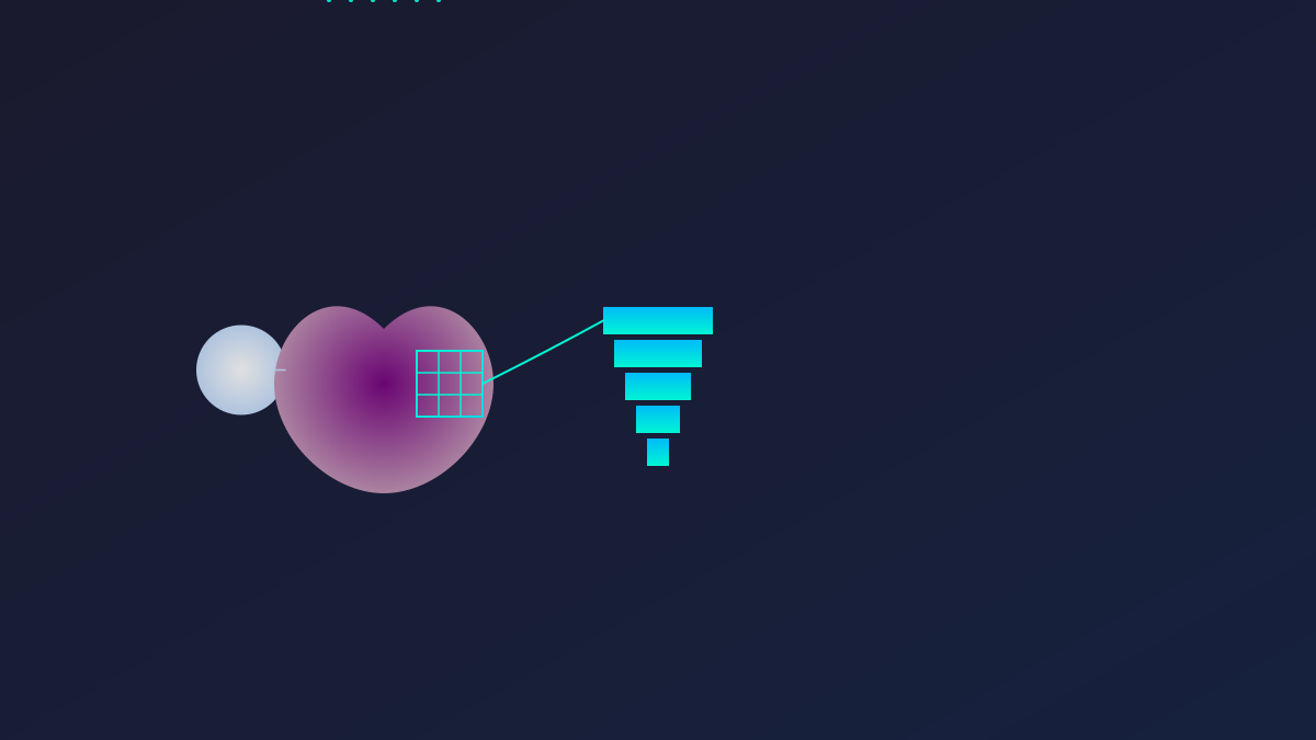
MRI image input → YOLOv8-OBB tumor localization → Tumor region extraction → EfficientNet-B0 classification → Output tumor type

Value of Oriented Bounding Box (OBB)

YOLOv8-OBB uses oriented bounding boxes, which fit the tumor shape more closely and reduce background interference. The annotation format is four corner coordinates.

Core Technology Stack

PyTorch, Ultralytics YOLOv8, OpenCV, NumPy, etc. Data preprocessing includes normalization, size adjustment, data augmentation, etc.

4

Section 04

Evidence: Dataset, Training Configuration, and Performance Evaluation Results

Dataset

Uses Kaggle's YOLO v11 Tumor Detection Dataset, with a total of 1947 MRI images (1362 for training / 395 for validation / 190 for testing), identifying four types of tumors (glioma, meningioma, pituitary tumor, no tumor).

Training Configuration

Input size: 224×224; Adam optimizer; learning rate: 0.001; batch size: 16; training for 5 epochs; using ImageNet pre-trained weights.

Performance

EfficientNet-B0 achieves a classification accuracy of 94.74%, precision of 95.34%, recall of 94.74%, F1 score of 94.90%; YOLOv8-OBB is evaluated via metrics like mAP and angle loss, and performs better than DenseNet121.

5

Section 05

Conclusion: Project Value and Gap from Clinical Deployment

This project demonstrates a typical application of deep learning in medical imaging: task decomposition (localization + classification), model selection (YOLO detection/EfficientNet classification), domain adaptation (transfer learning on medical datasets), and rigorous evaluation. The 94.74% accuracy is a good starting point, but to reach clinical deployment, larger datasets, strict validation, and regulatory approval are needed. The dual-model collaboration idea provides a reference template for medical AI.

6

Section 06

Suggestions and Future Directions: Clinical Application Prospects and Improvement Ideas

Clinical Significance

Can serve as an auxiliary tool for radiology: initial screening to mark suspicious areas, quantitative analysis of tumor size and location, and providing references for difficult cases.

Future Directions

  1. Larger-scale datasets; 2. Multimodal fusion (T1/T2/FLAIR sequences); 3. Pixel-level segmentation; 4. Clinical validation; 5. 3D MRI analysis.